Bee anatomy involves many names, some of which can be quite specialized.
Here is an illustration of the different structures so that you may find your way in the descriptions and identification criteria.
At the end, you will find a glossary in alphabetical order, if you are looking for a specific word.
Here is an illustration of the different structures so that you may find your way in the descriptions and identification criteria.
At the end, you will find a glossary in alphabetical order, if you are looking for a specific word.
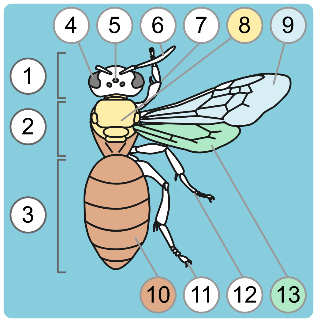
A. General traits: dorsal view of a bee.
Colored numbers refer to the entire colored area.
|
1. Head
2. Mesosoma (often called 'thorax' for simplicity) 3. Metasoma (often called 'abdomen' for simplicity) 4. Eye (= Compound Eye) 5. Anterior ocellus (Plural: ocelli) 6. Antenna (= Scapus + Pedicel + Flagellum) 7. Anterior leg (= Leg 1 = Foreleg) 8. Thorax (homologous to other insect's thorax) 9. Anterior wing (= Forewing) 10. Abdomen (homologous to other insect's abdomen) 11. Posterior leg (= Leg 3 = Hindleg) 12. Middle leg (= Leg 2 = Midleg) 13. Posterior wing (= Hindwing) Note that the thorax (A8) is only part of the Mesosoma (A2), as in other Apocrite wasps and ants. |
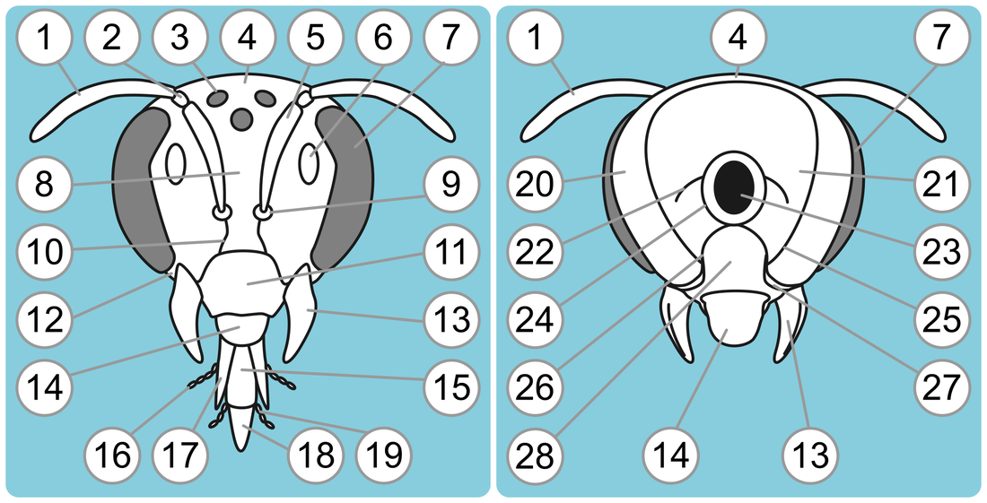
B. The head in frontal and posterior views
|
1. Flagellum (includes 10 or 11 flagellomeres, or antennal segments)
2. Pedicel (= antennal segment 2) 3. Posterior ocellus 4. Vertex 5. Scapus (= antennal segment 1) 6. Facial fovea 7. Eye (= Compound eye) 8. Frons 9. Antennal socket 10. Subantennal suture 11. Clypeus |
12. Malar space (= Malar area)
13. Mandible 14. Labrum 15. Prementum 16. Maxillary palpus (plural: palpi) 17. Galea 18. Glossa 19. Labial palpus (plural: palpi) 20. Gena 21. Occiput |
22. Occipital sulcus (= occipital carina)
23. Foramen magnum 24. Postocciput 25. Preoccipital carina (= Preoccipital ridge) 26. Hypostomal carina (= Hypostomal ridge) 27. Hypostoma 28. Proboscidial fossa |
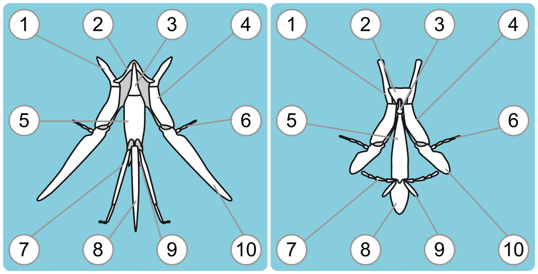
C. Mouthparts of long-tongue bees (Apidae+Megachilidae) and short-tongue bees (Andrenidae, Colletidae, Halictidae and Melittidae)
|
1. Cardo
2. Lorum 3. Mentum |
4. Stipe
5. Prementum 6. Maxillary palpus (plural: palpi) 7. Labial palpus (plural: palpi) |
8. Glossa
9. Paraglossa 10. Galea |
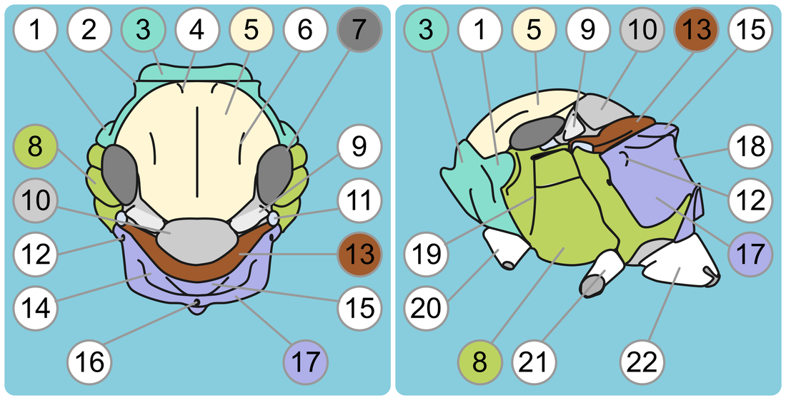
D. Mesosoma in dorsal and lateral (from the left) views.
Colored numbers refer to the entire colored area.
|
1. Pronotal lobe
2. Dorso lateral angle of the pronotum 3. Pronotum 4. Notaulus (plural: notauli) 5. Scutum (= Mesoscutum = Shield) 6. Parapsidal line 7. Tegula (plural: tegulae) 8. Mesepisternum |
9. Axilla (plural: axillae)
10. Scutellum 11. Insertion of the posterior wing 12. Propodeal spiracle 13. Metanotum 14. Dorsal part of the propodeum 15. Propodeal triangle |
16. Propodeal pit.
17. Propodeum 18. Posterior part of the propodeum 19. Episternal groove 20. Anterior coxa (= Procoxa = Coxa 1; plural: coxae) 21. Middle coxa (= Mesocoxa = Coxa 2) 22. Posterior coxa (= Metacoxa = Coxa 3) |
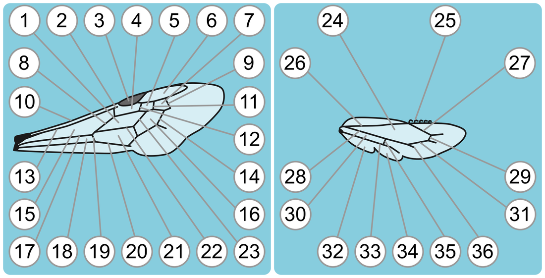
E. Anterior and posterior wings.
|
Anterior wing:
1. Medial cell 1 2. Prestigma (= 1st Abscissa of R1) 3. Pterostigma 4. Submarginal cell 1 5. Submarginal cell 3 6. Marginal cell 7. Submarginal cell 3 8. Basal vein (= Medial vein) 9. Submarginal crossvein 3 (= 2nd r-m crossvein) 10. Costal vein 11. Submarginal crossvein 2 (= 1st r-m crossvein) 12. Submarginal crossvein 1 (= 2nd abscissa of Rs) |
13. Radial vein (= Subcostal vein)
14. Recurrent vein 2 (= 2nd m-cu crossvein) 15. Radial cell 16. Medial cell 2 17. Medio-cubital vein (= M+Cu) 18. Cubital cell 1. 19. Cubito-anal crossvein (= cu-a = cu-v) 20. Anal vein (= Vannal vein = A = V) 21. Cubital cell 2 22. Cubital vein (= Cu) 23. Recurrent vein 1 (= 1st m-cu crossvein) |
Posterior wing:
24. Radial cell 25. Hamulus (plural: hamuli) 26. Radial vein (= R) 27. Radial Sector vein (= Rs) 28. Medio-cubital vein (= M+Cu) 29. Medio-radial crossvein (= r-m) 30. Cubital cell 31. Medial vein (= M) 32. Jugal lobe 33. Anal vein (= Vannal vein = A = V) 34. Anal lobe (= Vannal lobe) 35. Cubito-anal crossvein (= cu-a = cu-v) 36. Cubital vein |
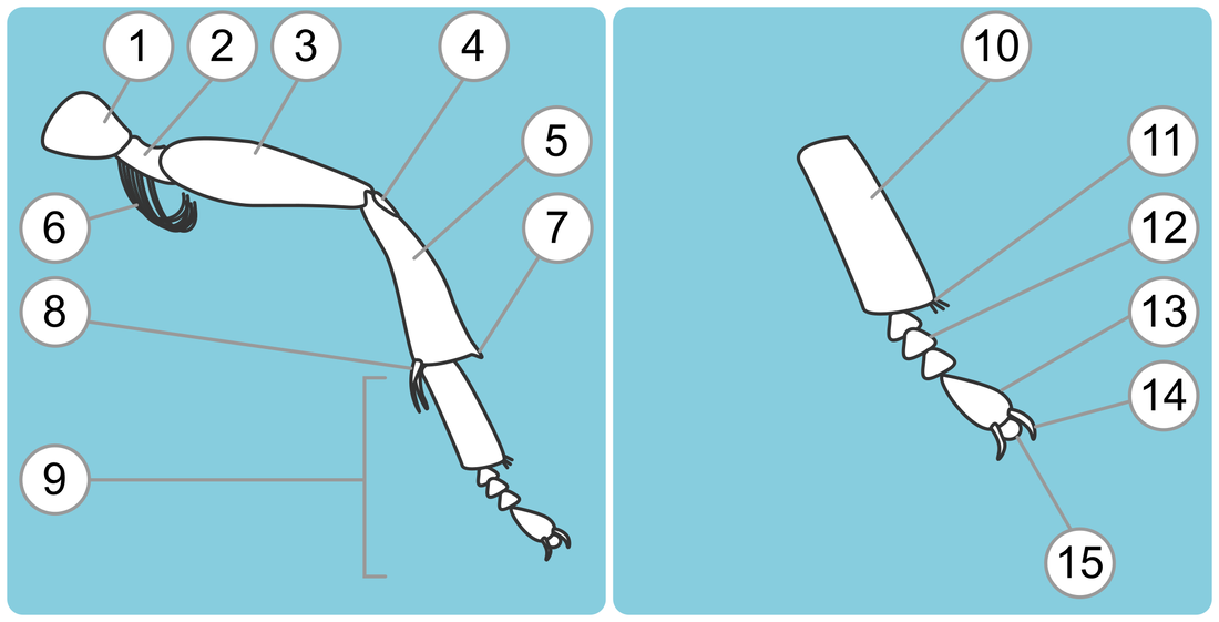
F. Posterior leg of a female bee and posterior tarsus
|
1. Coxa (plural: coxae)
2. Trochanter 3. Femur 4. Basitibial plate 5. Tibia |
6. Flocculus
7. Tibial spine 8. Tibial spur 9. Tarsus (plural: tarsi) 10. Basitarsus (= Tarsal segment 1 = Tarsomere 1) |
11. Penicillus
12. Mediotarsus (= Tarsal segment 3 (or 2 or 4) = Tarsomere 3 (or 2 or 4) ) 13. Distitarsus (= Last tarsal segment = Last tarsomere) 14. Tarsal claw 15. Arolium |
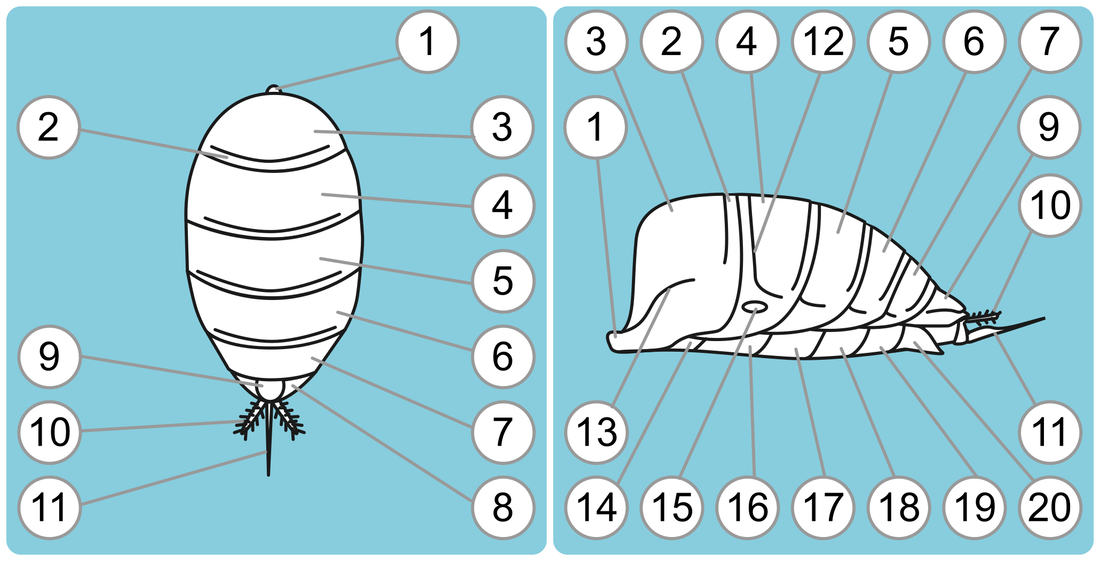
G. Metasoma of a female bee in dorsal and lateral (from the left) views
|
1. Petiole
2. Marginal area of T1 3. T1 (= Tergite 1) 4. T2 (= Tergite 2) 5. T3 (= Tergite 3) 6. T4 (= Tergite 4) 7. T5 (= Tergite 5) |
8. T6 (= Tergite 6)
9. Pygigial plate 10. Gonostylus 11. Stylus 12. Gradulus 13. Lateral line of T1 14. S1 (= Sternite 1) |
15. Lateral fovea
16. S2 (= Sternite 2) 17. S3 (= Sternite 3) 18. S4 (= Sternite 4) 19. S5 (= Sternite 5) 20. S6 (= Sternite 6) |
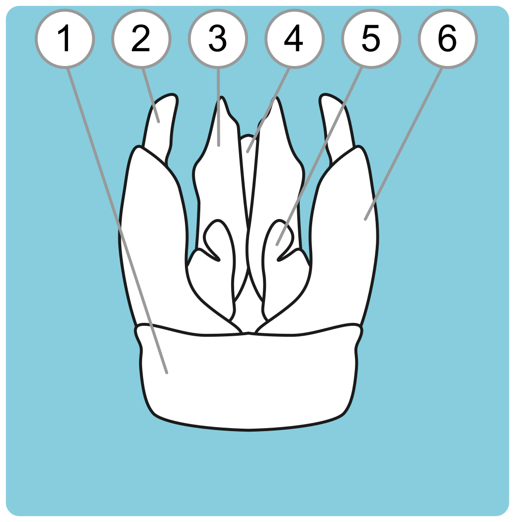
H. Male genitalia in dorsal view
Glossary
|
1st Abscissa of R1---E.2
1st m-cu (Crossvein)---E.23 1st r-m crossvein---E.11 2nd abscissa of Rs---E.12 2nd m-cu crossvein---E.14 2nd r-m crossvein---E.9 A (Vein)---E.20 A (Vein)---E.33 Abdomen (homologous to other insect's abdomen)---A.10 Abdomen (sensu Metasoma)---A.3 Anal lobe (= Vannal lobe)---E.34 Anal vein (= Vannal vein = A = V)---E.33 Anal vein (= Vannal vein = A = V)---E.20 Antenna (= Scapus + Pedicel + Flagellum)---A.6 Antennal segment 1---B.5 Antennal segment 2---B.2 Antennal socket---B.9 Anterior coxa (= Procoxa = Coxa 1; plural: coxae)---D.20 Anterior leg (= Leg 1 = Foreleg)---A.7 Anterior ocellus (Plural: ocelli)---A.5 Anterior wing (= Forewing)---A.9 Arolium---F.15 Axilla (plural: axillae)---D.9 Basal vein (= Medial vein)---E.8 Basitarsus (= Tarsal segment 1 = tarsomere 1)---F.10 Basitibial plate---F.4 Cardo---C.1 Clypeus---B.11 Compound Eye---A.4 Compound Eye---B.7 Costal vein---E.10 Coxa (plural: coxae)---F.1 Coxa 1---D.20 Coxa 2---D.21 Coxa 3---D.22 Cu (Vein)---E.22 cu-a (Crossvein)---E.19 cu-v (Crossvein)---E.19 Cubital cell---E.30 Cubital cell 1---E.18 Cubital cell 2---E.21 Cubital vein---E.36 Cubital vein (= Cu)---E.22 Cubito-anal crossvein (= cu-a = cu-v)---E.19 Cubito-anal crossvein (= cu-a = cu-v)---E.35 Distitarsus (= Last tarsal segment = last tarsomere)---F.13 Dorsal part of the propodeum---D.14 Dorso lateral angle of the pronotum---D.2 Episternal groove---D.19 Eye (= Compound Eye)---A.4 Eye (= Compound eye)---B.7 Facial fovea---B.6 Femur---F.3 Flagellum (includes 10 or 11 flagellomeres, or antennal segments)---B.1 Flocculus---F.6 Foramen magnum---B.23 Foreleg---A.7 Forewing---A.9 Frons---B.8 Galea---B.17 Galea---C.10 Gena---B.20 Glossa---B.18 Glossa---C.8 Gonobase---H.1 Gonocoxite---H.6 Gonostylus---G.10 Gonostylus---H.2 Gradulus---G.12 Hamulus (plural: hamuli)---E.25 Head---A.1 Hindleg---A.11 Hindwing---A.13 Hypostoma---B.27 Hypostomal carina (= Hypostomal ridge)---B.26 Hypostomal ridge---B.26 Insertion of the posterior wing---D.11 Jugal lobe---E.32 Labial palpus (plural: palpi)---B.19 Labial palpus (plural: palpi)---C.7 Labrum---B.14 Lateral fovea---G.15 Lateral line of T1---G.13 Leg 1---A.7 Leg 2---A.12 Leg 3---A.11 Lorum---C.2 M (Vein)---E.31 M + Cu (Vein)---E.17 M + Cu (Vein)---E.29 Malar area---B.12 Malar space (= Malar area)---B.12 Mandible---B.13 Marginal area of T1---G.2 Marginal cell---E.6 Maxillary palpus (plural: palpi)---B.16 Maxillary palpus (plural: palpi)---C.6 Medial cell 1---E.1 Medial cell 2---E.16 Medial vein (= M)---E.31 Medio-cubital vein (= M+Cu)---E.17 Medio-cubital vein (= M+Cu)---E.28 Medio-radial crossvein (= r-m)---E.29 Mediotarsus (= Tarsal segment 3 (or 2 or 4) = Tarsomere 3 (or 2 or 4) )---F.12 Mentum---C.3 Mesepisternum---D.8 Mesocoxa---D.21 Mesoscutum---D.5 Mesosoma (often called 'thorax' for simplicity)---A.2 Metacoxa---D.22 Metanotum---D.13 Metasoma (often called 'abdomen' for simplicity)---A.3 Middle coxa (= Mesocoxa = Coxa 2)---D.21 Middle leg (= Leg 2 = Midleg)---A.12 Midleg---A.12 |
Notaulus (plural: notauli)---D.4
Occipital carina---B.22 Occipital sulcus (= occipital carina)---B.22 Occiput---B.21 Paraglossa---C.9 Parapsidal line---D.6 Pedicel (= antennal segment 2)---B.2 Penicillus---F.11 Penis---H.4 Penis valve---H.3 Petiole---G.1 Posterior coxa (= Metacoxa = Coxa 3)---D.22 Posterior leg (= Leg 3 = Hindleg)---A.11 Posterior ocellus---B.3 Posterior part of the propodeum---D.18 Posterior wing (= Hindwing)---A.13 Postocciput---B.24 Prementum---B.15 Prementum---C.5 Preoccipital carina (= Preoccipital ridge)---B.25 Preoccipital ridge---B.25 Prestigma (= 1st Abscissa of R1)---E.2 Proboscidial fossa---B.28 Procoxa---D.20 Pronotal lobe---D.1 Pronotum---D.3 Propodeal pit---D.16 Propodeal spiracle---D.12 Propodeal triangle---D.15 Propodeum---D.17 Pterostigma---E.3 Pygigial plate---G.9 R (Vein)---E.26 R (Vein)---E.26 Radial cell---E.15 Radial cell---E.24 Radial Sector vein (= Rs)---E.27 Radial vein (= R)---E.26 Radial vein (= Subcostal vein)---E.13 Recurrent vein 1 (= 1st m-cu crossvein)---E.23 Recurrent vein 2 (= 2nd m-cu crossvein)---E.14 Rs (Vein)---E.28 S1 (= Sternite 1)---G.14 S2 (= Sternite 2)---G.16 S3 (= Sternite 3)---G.17 S4 (= Sternite 4)---G.18 S5 (= Sternite 5)---G.19 S6 (= Sternite 6)---G.20 Scapus (= antennal segment 1)---B.5 Scutellum---D.10 Scutum (= Mesoscutum = Shield)---D.5 Shield---D.5 Sternite---G.14,G.16-G.20 Stipe---C.4 Stylus---G.11 Subantennal suture---B.10 Subcostal vein---E.13 Submarginal cell 1---E.4 Submarginal cell 3---E.5 Submarginal cell 3---E.7 Submarginal crossvein 1 (= 2nd abscissa of Rs)---E.12 Submarginal crossvein 2 (= 1st r-m crossvein)---E.11 Submarginal crossvein 3 (= 2nd r-m crossvein)---E.9 T1 (= Tergite 1)---G.3 T2 (= Tergite 2)---G.4 T3 (= Tergite 3)---G.5 T4 (= Tergite 4)---G.6 T5 (= Tergite 5)---G.7 T6 (= Tergite 6)---G.8 Tarsal claw---F.14 Tarsomere (1)---F.10 Tarsomere (2,3 or 4)---F.12 Tarsomere (Last)---F.13 Tarsus (plural: tarsi)---F.9 Tegula (plural: tegulae)---D.7 Tergite---G.3-G.7 Thorax (homologous to other insect's thorax)---A.8 Thorax (sensu Mesosoma)---A.2 Tibia---F.5 Tibial spine---F.7 Tibial spur---F.8 Trochanter---F.2 V(Vein)---E.20 V(Vein)---E.33 Vannal vein---E.20 Vannal vein---E.33 Vertex---B.4 Volsella---H.5 |

